CT scan of small hearts could improve cardiovascular research
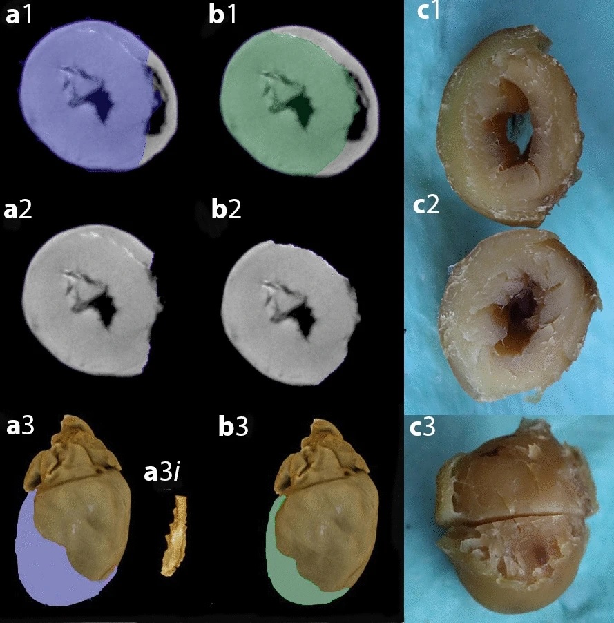
Micro-CT scan with virtual dissection of left ventricle is a non-destructive, reproducible alternative to dissection and weighing for left ventricular size. More information about this image is available here.
Use of laboratory mice for exploration into human disease is an essential activity that has led to many medical breakthroughs. It provides the foundational research needed to progress to human trials. In the case of cardiovascular disease, the ability to virtually dissect and examine the left ventricle in mice using CT scanning could be a leap forward for human cardiovascular research.
The left ventricle – one of the four chambers of the heart – is the strongest chamber, which pumps oxygen-rich blood to the body and creates blood pressure. The size and mass of the left ventricle provides important information about cardiovascular health. As we age or because of underlying medical conditions, the left ventricular size can vary. Its size is dependent on lifestyle choices, such as exercise, or medical conditions such as high blood pressure, valvular heart disease or genetically inherited conditions. If it increases significantly in size, it will result in cardiovascular problems.
In a world first study, Dr Ata Doost, ANU Medical School PhD Candidate, and colleagues have proven that micro-CT scanning (or virtual dissection) of the left ventricle in a mouse heart is as accurate as real tissue dissection in the laboratory. In the case of this study, real tissue dissection refers to the left ventricle being separated from the rest of the cardiac structure and physically weighed by a special scale.
The use of a virtual examination of the heart by CT scan image allows for the identification of any structural abnormalities such as left ventricular hypertrophy, congenital heart disease or abnormal communications between chambers that previously required fine cuts through the heart sample, staining and then examination under a microscope.
“Historically, laboratory tissue dissection for detailed examination of the heart has been a gold standard method in order to look for underlying normal and abnormal tissues or structures. These laboratory tests, however, are time-consuming, labour consuming and can result in irreversible tissue damage to the sample heart,” explained Dr Doost.
“CT-scan is a valuable tool to investigate organ structure in 3-dimensional micro-meter scale and helps us preserve the tissue for future dissection and examination if needed. This technique could be more accurate than laboratory dissection and weighing especially in small animal models. This study has shown that fundamental science researchers working on animal models of human illnesses can quickly, non-invasively, and less expensively examine small organs such as hearts using CT scanning.”